


Ever since its discovery over a century ago, x-ray has revolutionized every aspect of our lives. A computed tomography (CT) scanner is an x-ray based imaging system that is capable of providing non-invasive three-dimensional (3D) images, and has a wide range of applications in medical healthcare, industrial inspection, airline security, forensic investigation, scientific research, and many others.
Our research is typically focused at novel X-ray sources, detectors, signal processing, image reconstruction algorithms, as well as systems engineering. Below we showcase select research. Please see our publications page for a complete picture.
1. CNT X-ray Source Based Imaging
1.1 CNT Field Emission X-Ray Sources and Source Arrays
Carbon nanotube (CNT) x-ray source technology is viewed by many to be a disruptive technology that has the potential to fundamentally change how x-ray is generated and utilized. We have extensive experiences in developing this novel x-ray source technology. We invented and engineered a series of technologies/processes for reliably generating x-ray radiations from carbon nanotube field emitters. As a result, power/flux and lifetime of carbon nanotube x-ray sources were increased by more than 10-fold from previously reported values; both are critically important for in vivo biomedical imaging of small animals and ultrafast medical imaging of human subjects.

Paper (PDF): Guohua Cao, et al., “A dynamic micro-CT scanner based on a carbon nanotube field emission x-ray source”, Physics in Medicine and Biology 54, 2323-2340 (2009).
1.2 CNT X-ray Source Based Imaging Systems
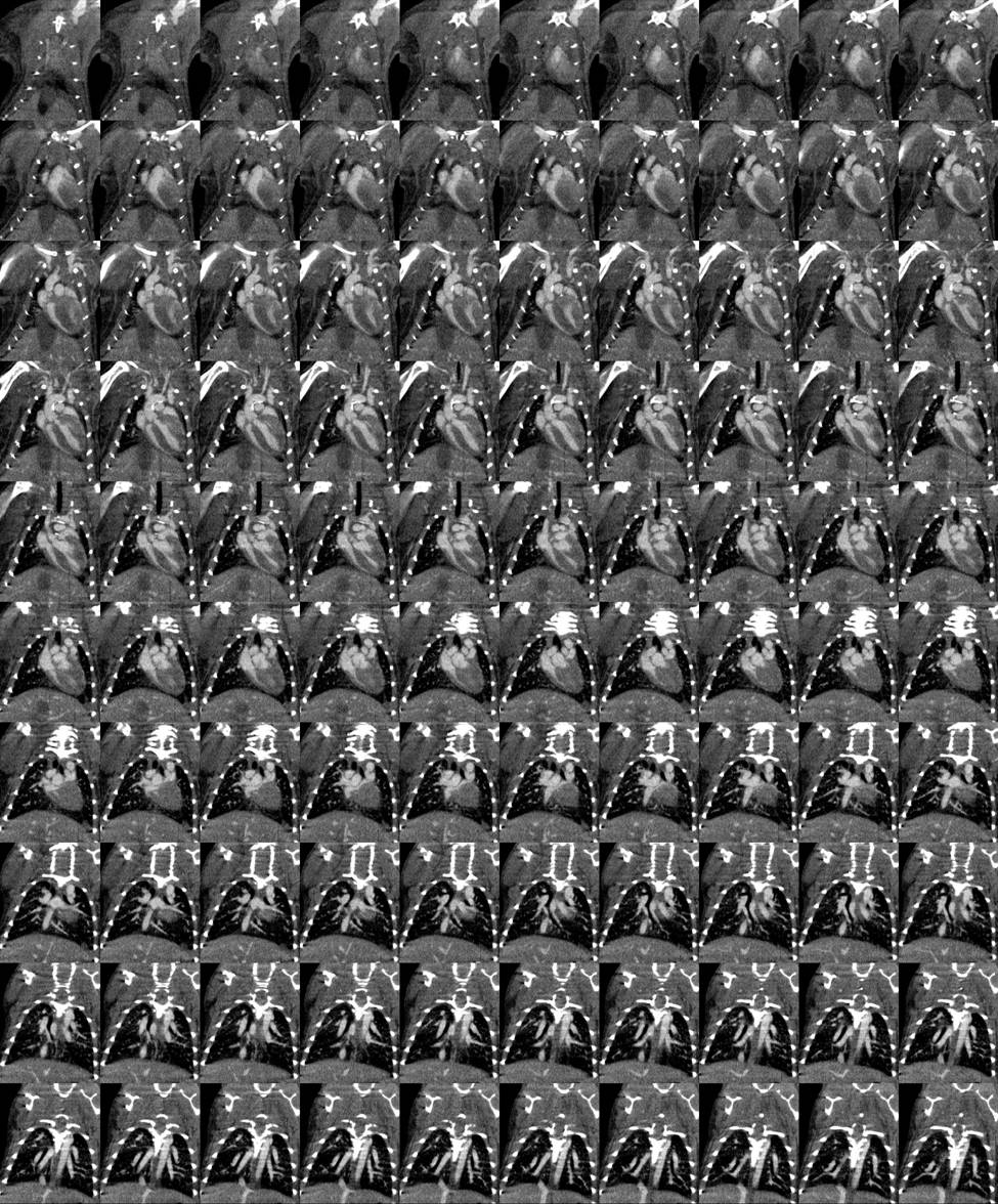
CNT Dynamic Micro-CT: The goal is to develop a dynamic micro-computed tomography (micro-CT) scanner with enhanced spatial and temporal resolutions, using the novel field emission micro-focus x-ray sources based on carbon nanotubes (CNT) field emitters. Here we utilize one of the unique advantages of a field-emission x-ray source: the ultrafast electronic switching and easy programming capability. Imaging sequence in a CNT dynamic micro-CT scanner can be readily synchronized to non-periodic physiological signals of free-breathing mice, hence dramatically reducing the physiological motion and producing much clearer delineation in morphology. Physiological functional information can also be obtained from imaging gated at the desired physiological phases.
Previously, we have successfully developed a prototype CNT dynamic micro-CT scanner and applied it to high-resolution imaging of various in vivo mouse models, including respiratory gated lung imaging, cardiac-and-respiratory gated cardiac imaging, atherosclerotic imaging, liver tumor imaging, as well as lung and colon cancer imaging. Recently, we further refined this CNT micro-CT to allow interior micro-CT imaging at greatly (up to 85%) reduced radiation dose. CNT interior micro-CT has been applied to image mouse hearts. [See some images at the gallery...]
We have brought this CNT dynamic micro-CT to a fully functional imaging device and shared the novel technology with the broad research community. Dr. Cao has built two CNT micro-CT scanners as the core imaging facilities in two institutions: one in the Biomedical Research Imaging Center at UNC-Chapel Hill; and the other in the Department of Radiology at University of Iowa - Carver College of Medicine.

Paper (PDF): Guohua Cao, et al., “Prospective-gated cardiac micro-CT imaging of free-breathing mice using carbon nanotube field emission x-ray”, Medical Physics 37, 5306-5312 (2010).
CNT Stationary Breast Tomosynthesis: One unique advantage of CNT x-ray source is its ability to easily package multiple x-ray pixels into one vacuum envelope to produce multi-beam x-ray source array. With multi-beam x-ray source x-ray, a spatially scanning x-ray beam can be easily generated via electronic activation of gate electrode at each x-ray pixel, thus enabling tomographic scanning without x-ray source rotation. A demonstration of such capability is a CNT Stationary Breast Tomosynthesis system developed at UNC-CH.
Paper (PDF): Guang Yang, et al., “Stationary digital breast tomosynthesis system with a multi-beam field emission x-ray source array”, SPIE-Medical Imaging 69131A-69131A-10 (2008).
2. New CT Architectures & Reconstruction Algorithms
In current CT architecture, both x-ray tubes and x-ray detectors are rotated mechanically around an object to collect large amounts of projection data. This architecture is not fast enough for patients with high or irregular heart rates. Furthermore, both x-ray beams and detectors of the current architecture are made wide enough so that the entire object is covered in the lateral direction without data truncation. Consequently, the high radiation dose from CT imaging remains a heightened public concern (especially for cardiac CT). To overcome these problems, we came out with an innovative stationary-sources CT architecture based on three stationary distributed x-ray sources and three smaller rotating x-ray detectors. Three source-detector chains can work in parallel to acquire three projections simultaneously and improve temporal resolution. Lower full-body radiation dose is expected for the proposed CT architecture because x-rays are restricted to irradiate a local smaller region. Combined with latest reconstruction algorithms for sparse-view CT, the proposed CT architecture will enable <=50ms temporal resolution and reduce radiation dose significantly.

a. Paper (PDF): Guohua Cao, et al., “A stationary-sources and rotating-detectors computed tomography architecture for higher temporal resolution and lower radiation dose” IEEE Access 2, 1263-1271 (2014).
b. Paper (PDF): Zhicheng Zhang, et al., “A Novel Sparse-View CT Reconstruction Method Based on Combination of DenseNet and Deconvolution”, IEEE Trans. on Medical Imaging 37(6), 1407-1417 (2018).
3. New Mechanisms for X-ray Imaging
Traditional x-ray imaging has a contrast mechanism based on the attenuation difference between different materials. It can provide high spatial resolutions to delineate morphology but is difficult in differentiating soft tissues that have clear differences in their chemistry, physical properties, and more generally functions. We have contributed to the advancement of the x-ray phase-contrast imaging and spectral imaging for enhanced soft-tissue contrast, and developed a new x-ray fluorescence imaging design for potential x-ray based molecular imaging.3.1 X-ray Phase Contrast Imaging
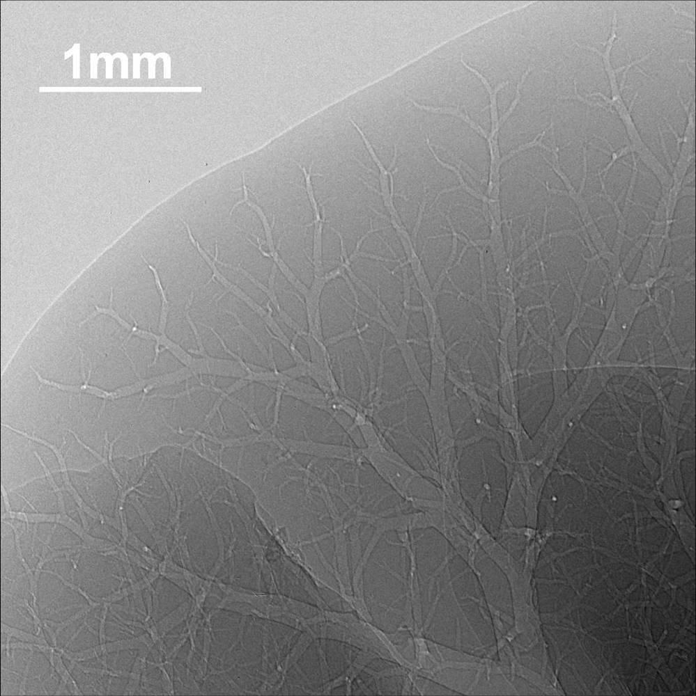
X-ray phase contrast imaging refers to using the phases shift introduced in an x-ray beam as it traverses a body to give contrast. Conventional x-ray imaging does not show the effects of the deflection of the trajectories of the x-rays in the images because of the large size of the x-ray source. Major advantage of phase contrast imaging over conventional imaging is enhanced image quality at reduced radiation dose.
We have built an x-ray phase contrast imaging system using a micro-focus x-ray source and a high-resolution CCD camera arranged in high magnification geometry such that small features down to 10 microns can be resolved. With this system, we have investigated x-ray phase contrast imaging of technical samples, simulated breast phantoms, and actual malignant biological tissues. We are actively investigating some new phase retreival methods such as structured illumination based method and physics-model based computation method. Our goal is to develop and engineer a compact x-ray phase contrast imaging system suitable for clinical applications.

Paper (PDF): Guohua Cao, et al., “X-ray phase contrast imaging: Transmission functions separable in cylindrical coordinates”, J. Appl. Phys. 105, 102002 (2009).
3.2 X-ray Fluorescence Molecular Imaging
X-ray Fluorescence Molecular Imaging (XFMI) has shown great promise as a low-cost molecular imaging modality for clinical and pre-clinical applications with high sensitivity. Recently, progress has been made in enabling the XFMI technique with laboratory x-ray sources for various biomedical applications. However, the sensitivity of XFMI still needs to be improved for in vivo biomedical applications at a reasonably low radiation dose. In laboratory x-ray source based XFMI, a main factor that limits the molecular sensitivity of XFMI is the scatter x-rays that coincide with the fluorescence x-rays from the targeted material. We experimentally investigated the effects of excitation beam spectrum on the molecular sensitivity of XFMI, by quantitatively deriving Minimum Detectable Concentration (MDC) under a fixed surface entrance dose at three different excitation beam spectra. Our result shows that the MDC can be readily increased by a factor of 5.26 via excitation spectrum optimization. We also developed a numerical model and validated it with our experiment data. Our findings from could find applications for in vivo pre-clinical small-animal XFMI in the future.
Paper (PDF): Xu Dong, et al., "Improving Molecular Sensitivity in X-Ray Fluorescence Molecular Imaging (XFMI) of Iodine Distribution in Mouse-Sized Phantoms via Excitation Spectrum Optimization". IEEE Access 6: p. 56966-56976 (2018).
3.3 Spectral X-ray Imaging
Spectral x-ray imaging leverages the energy-sensitive information during x-ray image acquisition, and thus can provide x-ray attenuation coefficients of materials at multiple x-ray energy bins. It holds a great potential for material identification via calculation of effective atomic numbers and effective electron densities. We developed a deep-learning (DL) based method, which is more accurate for calculation of effective atomic numbers and effective electron densities from spectral x-ray images. This can lead to more accurate material identification, which can find use in improved tissue identification in biomedical imaging.
Paper (PDF): Xu Dong, et al., "An improved physics model for multi-material identification in photon counting CT". Medical Imaging 2019: Physics of Medical Imaging, 10948: p. 109484O International Society for Optics and Photonics (2019).